
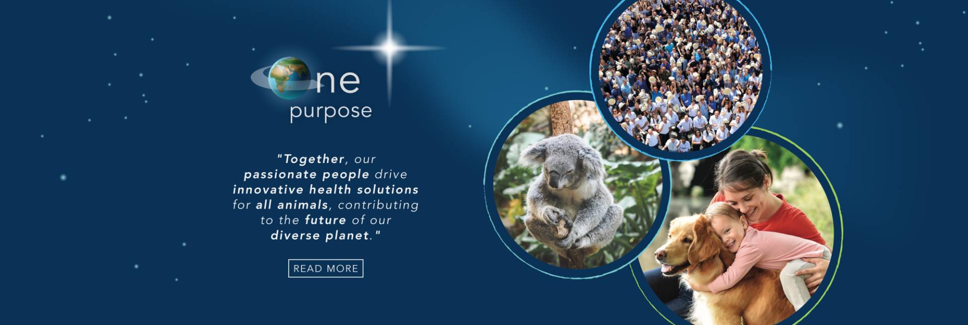
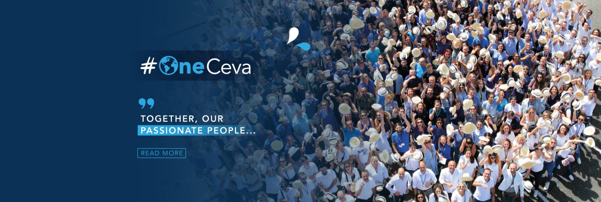
Together, our passionate people...
Our #OneCeva community is the driving force behind everything we undertake.
We build our business together, with our colleagues, with our customers and partners who put their trust in us every day.
Our #OneCeva community is the driving force behind everything we undertake.
We build our business together, with our colleagues, with our customers and partners who put their trust in us every day.

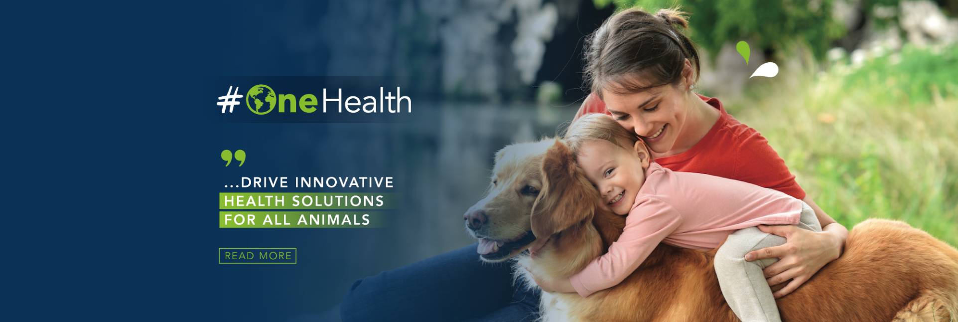
Ceva strives to be more than just a company committed to animal health, but a One Health company that plays an essential role in preserving the health of all: from animals to humans to our planet.

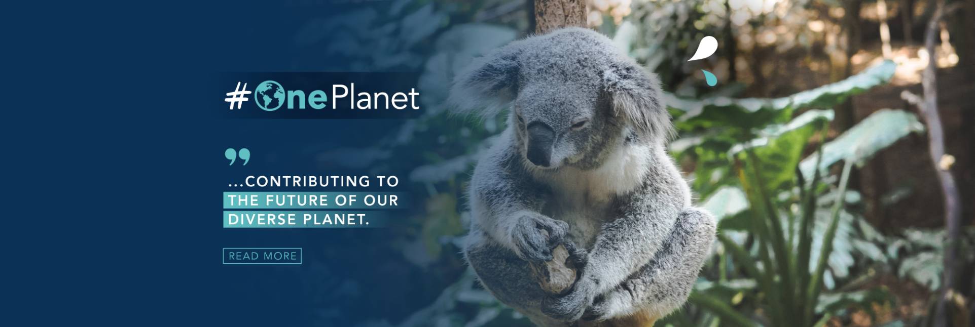
At Ceva, we are committed to preserving the diversity of our planet.
We understand the crucial importance of protecting our environment to ensure a sustainable future for all.
World Animal Vaccination Day: Ceva Santé Animale emphasizes the benefits of preventive medicine
One Health champion Dr Simon Doherty is recognised as “Veterinarian of the Year 2024” of the WVA Global Veterinary Awards supported by Ceva
Are you ready to join us? Ceva offers much more than a job, it is a life experience ...